Diseases of Uvea for Medical Students: Let’s start with Opthalmology with its basic Diseases of Uvea. I made this blog useful mainly for undergraduate medical students and those who are preparing for NExT, PLAB, and USMLE exams.
First, there will be clear-cut exam-oriented points on Diseases of Uvea notes for Medical Students then at the bottom there will be a link to the Free online MCQs on Diseases of Uvea and these MCQs are specially made up for NExT, PLAB, and USMLE exams. Jump to,
Page Contents
ToggleDiseases of Uvea for Medical Students
Uveitis
Inflammation of uveal tract
- Iris – Iritis
- Choroid – Choroiditis
- Ciliary body – cyclitis
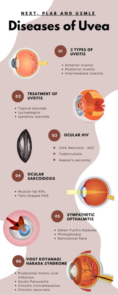
Classification
Anterior Uveitis
- Iritis
- Iridocyclitis (only Pars Plicata of CB) – 1st most common among Uveitis
Intermediate Uveitis
- Pars planitis
- Vitritis
Posterior Uveitis – 2nd most common
- Choroiditis
Panuveitis
- Sympathetic Ophthalmitis
- Vogt Koyanagi Harada Syndrome
Other classifications
- Acute Uveitis
- Chronic Uveitis (Inflammation more than 3 months)
- Granulomatous Uveitis – (TB, Syphilis, Sarcoidosis ).
- Non granulomatous Uveitis
One must right all these things in the diagnosis. For Eg Acute Anterior Nongranulomatous Uveitis.
Anterior Uveitis
Causes
- M/C – Idiopathic ( 50% )
- HLB B-27 Spondyloarthropathies
- Ankylosing spondylitis ‘ FlipFlop‘ – Young male with low back pain with red painful eyes.
- Inflammatory Bowel Disease – Ulcerative colitis & Crohn’s disease
- Psoriatic Arthritis
- Reiter’s syndrome (Reactive Arthritis) – Triad of Conjunctivitis, Urethritis, and Arthritis
- JRA – Juvenile Idiopathic Arthritis
- Children < 16 (Particularly girls )
- Pauciarticular, ANA positive, RF negative
- C/ I to IOL
- White eye Uveitis
- Arthritis – Uveitis – Cataract
Symptoms
Acute painful, red eye with loss of vision.
Signs
- Circum ciliary congestion (circumcorneal congestion)
- Bluish red,
- Radial rays
- Anterior ciliary vessels congestion
- Cells – Neutrophils ( WBC’s in the AC
- The sign of Active Uveitis
- The earliest sign of Active Uveitis
- Flare – Severe uveitis
- Turbid aqueous humor due to serum proteins from Ureal vessels
- Keratic precipitates
- Presence Cornea of N & L in the inferior cornea in the shape of a triangle (Arlt’s triangle)
- Mutton-feetKPs (Granulomatous Uveitis)
- Iris Nodules
- Koppe’s (Pupillary margin) and Busacca’s nodules (Surface of Iris) – Granulomatous Uveitis
- Synechiae
- Iris attached to Cornea – Anterior synechiae – Because Angle blocked (No drainage of AH) – Leads to glaucoma
- Iris attached to Lens – Posterior synechiae – Leads to cataract
- Miosis
- LOW TOP – Inflammed CB- Does not produce AH
- Festooned Pupil – Anterior uveitis causing posterior synechiae
- Hypopyon – Collection of pus in AC
Intermediate Uveitis
Rare, chronic, relapsing
Causes
- Idiopathic (70%) – Pars Planitis
- Sarcoidosis
- Multiple sclerosis
Symptoms
- Floaters / Muscae (Black spots surroundings)
- Neutrophils and WBC in Vitreous Humour
- Myopic and Vitreoretinal pathology
- Cystoid Macular Edema
- Snowballs and snow banks
Posterior Uveitis
Choroiditis
Infection Causes
- Toxoplasmosis – From cats
- Tuberculosis
- Toxocariasis
- CMV, Herpes, HIV
Autoimmune Causes
- Sarcoidosis
- Poly Arteritis Nodosa
- Scleroderma
Symptoms
- Chorio retinitis
- Vasculitis of retinal vessels
- Vitritis
Signs:
- Creamy yellow patches
- CR, Vasculitis vitritis (headlight and fog )
Treatment for Uveities
For Anterior Uveitis
Topical steroids – DOC of AU
No degradation of ECM on Trabecular meshwork leads to Biological edema and causes Glaucoma
So more powerful steroid leads to more glaucoma
Dexamethazone ↑ glaucoma
Fluromethazone ↓ glaucoma
Cycloplegic – DOC for Acute Uveitis
- Relaxes the ciliary spasm
- Dilates the pupils and breaks the synechiae
- Reduce vascularity of CB
Atropine ointments – 14 days – in children DOC (as Powerful ciliarytone seen in children)
Homatropine -3 days (Used)
Cyclopentolate – 1 day (Mostly used)
Tropicamide – 6 hours
For Intermediate Uveitis
Steroid injections – Triamcinolone
- subconjunctival
- subtenon’s
No role for cycloplegics in Intermediate Uveitis as there is no pain and no posterior synechiae to break
For Posterior Uveitis
Antimicrobials for infectious disease
Spiramycin DOC For toxoplasmosis in pregnancy.
TB – ATT
CMV – Grancyclovir
HIV – HAART therapy
Systemic steroids for non-infection (3 months only) cause S/E moon face.
Fuch's Heterochromic Iridocyclitis
Idiopathic, chronic, unilateral, low-grade Anterior Uveitis
Rubella virus cause
- Heterochromia
- cataract
- Diffuse, stellate KPs (Herpetic Iridocyclitis)
Minimal symptoms – Floaters, discovered when loss of vision – cataract, Posterior synechiae never present
Amsler’s sign – Paracentesis induces Hyphema
Topical/systemic steroids should be avoided (S/ E – Glaucoma)
Vision-threatening complications
- Cataract
- Glaucoma,
- Vitreous opacification.
Posner Schlossman Syndrome/ Glaucomatocyclitic Crisis
- Rare, recurrent, unilateral attacks of uveitis in young to middle-aged males
- Mild attacks of uveitis with minimal flare, white KPs- markedly out of proportion to the uveitis
- ↑ IOP – Corneal edema – mild loss of vision.
- Between attacks, the eye is normal.
- Association with CMV is seen.
- Anti glaucoma drugs, tropical steroids.
Ocular Manifestations in HIV
Commonest manifestation – Retinal microangiopathy – cotton wool spots (HT &DM), hemorrhages, microaneurysms
Commonest ocular infection – CMV retinitis
Commonest systemic infection – Tuberculosis
Commonest tumor – Kaposi’s sarcoma
Lymphoma
OSSN – Ocular Surface Squamous Neoplasia
CMV Retinitis
M/C cause of blindness in AIDS
CD < 50 cells /ml
CMV Retinitis incidence: 30% in pre HAART < 5% in HAART
Floaters, Photopsiae, loss of vision
3 patterns – Pizza pie, Brush Fire, Frosted branch angiitis.
DOC – HAART, Oral valgan cyclovir
Immune Recovery Uveitis
Para doxical worsening of intraocular inflammation- improved immunity HAART
M/C complication of HAART
The immune system recovers – attacks CMV – ocular inflammation prominent in vitreous
Risk factors – CD4 count 100 cells 1μL, IV cidofovir
- Floaters,
- Photopsiae,
- Redress,
- Pain,
- Posterior synechice,
- Hypopyon,
- IRV between
- cataract,
- vitriis,
- Optic disc edema
- EpiRetinal membrane.
Rx =) Periocular and intravitreal steroids.
Opportunistic Infections of Eye
2 Bacteria – TB, Syphilis
2 Fungus – cryptococcus, candida
2 Viral – Herpes zoster, CMV
2 Parasite – Pneumocystis, Toxoplasmosis
Ocular Sarcoidosis
25% sarcoidosis have ocalar involvement
Colored races I scan dinavians
A/I/P/ Pan Uveitis
Bilateral, granulomatous, chronic uveitis
7 ocular signs
- Mutton Fat KRS
- Tent shaped PAS/Berlin nodules In TM
- Vitreous opacities – String of Pearl
- Candle wax drippings (Tache de bougie)
Chest x-ray – Best investigation.
Elevated ACE and soluble IL 2 receptors.
The topical steroid with cycloplegic
M/C cause of Loss of Vision of cystoid macular edema
Sympathetic ophthalmitis
Bilateral, granulomatous, pan uveitis
Trauma (Penetrating/perforating injury)
CB injury – Uveal pigment – Act as FB (Antigen) – Auto Ab produced – Granulomatous Inflammation (Uveitis ) – Sympathetic ophthalmitis – another eye
Metallic Foreign body
Injury to CB a maximum – amount of Sympathetic Opthalmitis within 2 weeks to 2 months
Dalen Fuch’s Nodules – lymphocytes in the macula
M/C in children
Earliest Symptom
Photophobia
Accomodation failure (loss of NV)
Earliest Sign
Retrolental flare
Mutton fat KPs
Hypotony
Treatment
Enucleation within 14 days of trauma
Repair the injured eye
Systemic steroids
PL -ive, 6/6
6/60, 6/6
PL -ive, HM (+)
Vogt Koyanagi Harada syndrome
Bilateral, granulomatous, panuveitis
Neurological /auditory in volvement in Pigmented races in absence of ocular trauma or surgery
Young famales 20- 50
Symptoms
Prodromal mimics viral infection, neck stiffness, infection, hearing loss, headaches, tinnitus, vertigo
Acute – Panuveitis with serous retinal detachments.
Chronic convalescence – Poliosis, vitiligo, alopecia, depigmentation of choroid – sunset glow.
Chronic recurrent – recurrent granulomatous Anterior uveitis, mutton fats-kPs.
Ocular signs
B/L Panuveitis / Posterior Uveitis with multifocal choroiditis – serous Retinal Detachments – optic disc Hyperemia & edema
Perilimbal vitiligo – Sugiura’s sign
Treatment – High dose systemic steroids
TAKE FREE MCQs on Diseases of Uvea
I’m Nawin (Admin), a seasoned doctor and accomplished content writer with 8 years of experience. Join me as I unravel the latest breaking news, unveil behind-the-scenes happenings, and explore the aftermath scenes. With my expertise, I’ve crafted this renowned news site to provide you with an authentic perspective on daily happenings. Get ready to delve into a world of truth and knowledge on Medico Topics.